Cell Biology: What role does the Spir-Capu complex play in oogenesis?
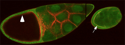
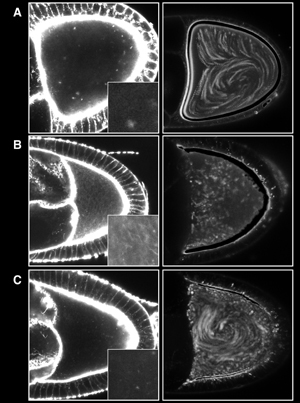
Spir and Capu are functionally linked to both the actin and microtubule cytoskeletons in Drosophila oogenesis. Both anterior-posterior and dorsal-ventral axes are established in the oocyte. In spir and capu mutants, axis determination fails and the timing of a dramatic process, microtubule-based streaming, is disrupted. I found that Spir is localized at the oocyte cortex through mid-oogenesis but is absent precisely when streaming normally begins (Fig. 3). An actin mesh, likely built by Spir and Capu, was recently described that is implicated important for streaming regulation (Fig. 4). In addition, Capu binds and bundles actin filaments and microtubules. Spir competes with this binding activity.
We are testing the roles of Spir and Capu by introducing rationally designed mutations and truncations of Spir and Capu based on our biochemical data. For example, we will determine whether actin nucleation, microtubule binding and/or Spir-Capu interaction are essential to polarity determination in oogenesis. The ability to combine genetic, cell biological, and biochemical approaches in one system makes Drosophila oogenesis an excellent model for studying cytoskeletal regulation and cell polarity. |

Immunofluorescence image of stage 9 and 10 Drosophila egg chambers. Spir (green) colocalizes with filamentous actin (red) at the oocyte cortex (arrow) only before stage 10 (arrowhead).

Correlation of actin mesh and cytoplasmic streaming. The left column is images of actin in ooctyes, stained with AlexaFluor488-phalloidin. Insets are 4x. The right column is overlays (thirty images acquired every ten seconds) of trypan blue stained yolk granules. (A) Stage 9, wild type. (B) Stage 10b, wild type. (C) Stage 9, capu null mutants.
|