Molecular Mechanism: How do Spir and Capu collaborate?
Studying the collaborative nucleation mechanism of the Spir-Capu complex is critical to understanding its cellular role. We recently learned that Spir and Capu bind directly to each other with high affinity in a 1:1 complex. Based on these studies, on our understanding of how Spir nucleates actin (movie,right) and a great deal of data about other formins, we have developed a working model, in which Spir plays the role of actin nucleator and Capu, protects the fast-growing barbed end of nascent filaments from being capped (see below). To test this model we need to know if Spir and Capu remain associated upon nucleation. The two alternate mechanisms have distinct structural and functional consequences because they result in filaments of opposite polarity at the membrane. Single molecule fluorescence microscopy will provide a powerful way to study the complex, enabling us to directly observe the behavior of the Spir-Capu complex with respect to the actin filaments they nucleate.
Because Spir has a modified FYVE zinc finger (mFYVE in figure below), Spir is depicted as a membrane-associated dimer (C, below). Classical FYVE domains are dimers that bind PI(3)P. The FYVE domain in Spir has a large insert and is missing conserved residues that determine lipid specificity. We need to perform structural studies and lipid binding assays to characterize this atypical FYVE domain.
Mechanism of actin nucleation by Spir
Left: Domain organization of Spire and Cappuccino. Right: Model of nucleation by the Spir- Capu complex
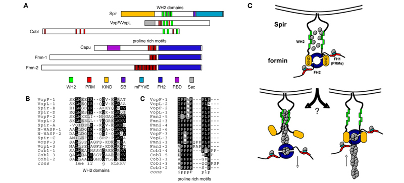
(A-B) Domain organization of Spir- and Capu-family. Top: The central region of Spir proteins contains a cluster of four actin-binding WH2 motifs, which nucleate actin. The C-terminal part consists of a modified FYVE zinc finger (mFYVE), which targets the protein to intracellular membranes. The adjacent Spir-box (S-box) is similar to motifs found in proteins that bind Rab-3a and may also play a role in subcellular localization. The KIND domain is a novel motif that may function as a protein-protein interaction module. Bottom: Capu, a formin, contains a proline rich region, the Formin Homology-1 domain (FH1), and a C-terminal Formin Homology-2 domain (FH2) that dimerizes and nucleates actin and a putative Rho-binding domain (RBD).
(C) Model of nucleation by the Spir- Capu complex. Actin orientation with respect to the membrane depends on Spir-Capu interaction. (Arrow indicates direction of rapid growth.)