Mammalian Disease: Is the Spir-Capu complex a polarity factor in other cell types?
Very little is known about Spir and Capu in species other than Drosophila. Genomic evidence and expression data suggest that the Spir-Capu complex is conserved throughout metazoans. In contrast to Drosophila, mammals have two isoforms of both Spir and Capu. One pair is expressed in epithelial cells, the second pair in neurons. Notably, both are polar cell types. Epithelial cells form a crucial barrier between organs and their environments. Proper polarity is essential to their function: polarity loss is a hallmark of some cancers and itself can lead to serious diseases such as cystic fibrosis. The Capu homolog, Formin-1 is localized in adherens junctions, structures that define different domains within epithelial cells. We don’t know if Spir and Capu function as a complex at adherens junctions or elsewhere in mammalian cells. Other evidence implicates Spir in transport pathways, interacting with Capu and Rab11 at the TGN and recycling endosomes (Fig. 6).
To understand how mammals utilize multiple isoforms of Spir and Capu, we need to determine whether or not the two isoforms are biochemically distinct, are differentially regulated or are only expressed in different cell types. The answers to these questions will provide insight into the function of the Spir-Capu complex and fundamental questions about the actin cytoskeleton and cell polarity. Understanding the regulation of a system necessary for female fertility and epithelial polarity could lead to sought after diagnosis and treatment.
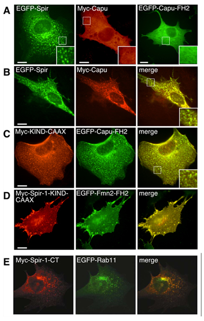
Right: NIH 3T3 cells transfected with individual expression vectors (A) or cotransfected with two expression vectors (B-E) encoding the indicated proteins. EGFP fusion proteins are green and the Myc-tagged counterpart is localized by immunofluorescence using anti-Myc antibodies (red). (A) When expressed alone, full length Capu and Capu-FH2 are diffuse throughout the cell. Spir is punctate. (B) When cotransfected with Spir, the localization of Capu shifts to a punctate pattern coinciding with Spir. (C) The Spir-KIND domain and Capu-FH2 domain are sufficient for colocalization. The KIND domain is driven to membranes by a CAAX box. Capu-FH2 is found concentrated at these same structures. (D) These interactions are conserved in mammalian proteins. Scale bars are 10 mm. Insets are expanded 2.3 times. (E) The C-terminal half of mammalian Spir-1 colocalizes with Rab11. (E is from Kerkhoff et al., Current Biology 2001) |